ARCHIVE - ARTICLES 2011

February 2011 - Laminitis
February 2011 - Mediolateral hoof balance in relation to the handedness of apprentice farriers
February 2011 - Nutrition and White Line Disease
![]() Archive - Forge Magazine - February 2011
Archive - Forge Magazine - February 2011
LAMINITIS
By Jim Ferrie FWCF
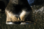
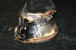
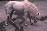 An actively laminitic horse
Laminitis is a disease associated with ischaemia (a restriction in blood supply that leads to damage or dysfunction of tissue) of the digital epidermal tissues. The bond between the dermal and epidermal laminae (the interlaminar bond) is the only means of support of the coffin bone within the hoof. Horses are suspended within their hooves by this interlaminar bond and if the laminar bond is destroyed; the pedal bone moves distally within the hoof capsule.
Laminitis can be acute (actively laminitic) or chronic (has suffered from laminitis in the past but is not painful presently).
Laminitis can be rotational (the laminar bond has broken down at the dorsal region of the hoof and the coffin bone is pulled downwards by the deep flexor tendon) or sinking (where a greater proportion of the laminar bed is deprived of blood, and, consequently nutrients, resulting in the coffin bone sinking downwards within the hoof capsule).
Common causes
Obesity
Laminitis caused by obesity is the most common type seen in the UK. Many horses and ponies do little work and unless calorie controlled diets are provided, these animals easily become overweight. Native ponies and cobs are very efficient at metabolising low energy forage and are unable to cope with fertilised cattle pasture.
Toxaemia
Toxic conditions such as metritis (retained afterbirth) put mares at high risk of developing laminitis.
Mechanical
Fast or prolonged work on hard surfaces may induce laminitis, and, following a non-weight bearing lameness, the contralateral limb may founder.
Hormonal
Some cases of laminitis appear to be thyroid problem related.
Cushing's disease
Symptoms of Cushing's disease in elderly animals include an enlarged pituitary gland or a pituitary tumour, which can cause the adrenal gland to release abnormal amounts of cortisol into the bloodstream (cortisol is a steroid hormone that is released in response to stress and leads to an increase in circulating blood sugar). Increased levels of cortisol often leads to the animal developing laminitis.
Diabetes
Variations in insulin levels often seen in diabetes (metabolic syndrome) are thought to induce laminitis.
Stress
Stress, such as overworking unfit horses, prolonged travelling in hot (or cold) conditions, may result in laminitis in some animals.
Heat in the feet; is a very unreliable diagnostic indicator. Foot temperature normally varies throughout the day.
The downward force of the bodyweight is neutralised by the resisting force of the ground. The primary forces involved are the pulling force of the deep digital flexor tendon, and the tearing force at the junction of the white line and the sole.
Biomechanical forces
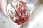
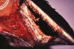 Morbid specimen showing total irreparable laminar damage
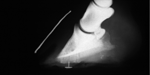
 Venogram showing no blood supply to the dorsal laminae
Treatment
Laminitis is always life threatening, there is no such thing as a 'touch' of laminitis. Therefore veterinary advice should be sought immediately. Usually, non-steroidal anti-inflammatory drugs (NSAIDs) will be prescribed, in conjunction with frog support, and removal of the cause (if it is known).
Farriery treatment normally follows the abatement of the symptoms, generally when the animal’s comfort level allows a hoof to be lifted without too much discomfort to the opposite limb, and radiography is advisable to locate the position of the bones inside the hoof capsule.
A heart bar is my shoe of choice, usually made of steel or an alloy, and nailed on lightly. This shoe offers stability to the bone column of the leg and hoof.
New technology allows us to glue hydroplastic heart bars to feet, should nailing prove too traumatic.
Hereditary predisposition to laminitis in this country is unproven. However, families of animals often have the same owner whose predisposition to recurring poor management is certainly proven.
Laminitis does not just affect the front feet, although because 65% of bodyweight is carried on the forelimbs, the front feet are often more badly affected.
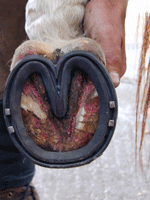 Concave heartbar shoe
 Loaded heartbar
 X ray showing seroma and sinking
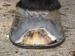 Dry dorsal seroma
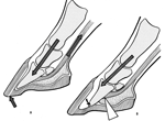
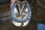
Fitting a heartbar shoe to a laminitic horse
The correct method of application of a heartbar shoe is crucial to the recovery of a laminitic animal. This should always be done with veterinary consultation.
Recent x-rays are essential to help position the shoe correctly.
The shoe is designed so that the frog carries a significant proportion of weight that would have been normally supported by the wall and sole; especially at the heels. The farriery term for this is a loaded heartbar shoe.
The bar in the shoe replicates the triangular shape of the frog, and lays flat on it within its perimeter. The tip of the bar must be perpendicular to a line drawn from the extensor process of the coffin bone within the hoof capsule The drawing pin shows the true point of the frog, giving an external reference point.
If the bar in the shoe is positioned too far forwards, the arterial blood supply to the sole will be compromised usually resulting in an abcess. If it is positioned too far back, the tip of the bone is driven downwards and can protrude through the sole.
The x-rays will not only show the current position of the coffin bone in relation to the hoof capsule, but can also show any pockets of gas which have filled the void created by the dead laminae.
When fitting a loaded heartbar, any serum or gas pockets should be opened to release the hydrostatic pressure within the hoof and this offers the horse some relief.
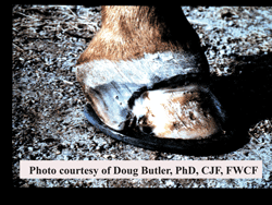
It should be remembered that this breakdown of the laminar bond occurs in three dimensions. Often the bone will displace medially due to the centre of weight bearing being carried down the inside of the limb.
 The tip of the coffin bone appearing through the side
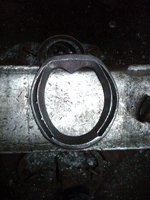
Right front hoof showing medial displacement of the hoof capsule
Note the growth rings wider on the lateral side
Shoeing the chronic laminitic horse
As soon as turning in a tight circle is not painful, heartbars may be removed and replaced with shoes that don’t impede the distended laminae at the toe – either unclipped or side clipped shoes.
The prognosis of a full recovery is always guarded, however, if the source can be found and eliminated or managed, then it is not unreasonable to expect a return to limited work if regular farriery and veterinary management is provided.
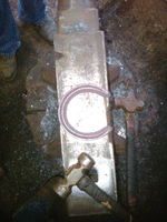
 Shoe placement on a chronic laminitic
This article appears on www.j-aferrie.com and is reprinted with permission of the author.
![]() Archive - Forge Magazine - February 2011
Archive - Forge Magazine - February 2011
Mediolateral hoof balance in relation to the handedness of apprentice farriers
A. Ronchetti, P. Day, R. Weller of the Royal Veterinary College, UK.
E-mail for correspondence: [email protected]
In a recent study carried out at the Royal Veterinary College, London, and published in Veterinary Record, (Vol 168, Jan 15, 2011, p 48) it was found that there was difference in mediolateral hoof balance for each forelimb, depending on whether a farrier was right-handed or left-handed. Studies of visuospatial awareness and, in particular, brain hemisphericity and line bisection have shown that there is a significant difference in the ability of right-handed and left-handed people to determine a midpoint accurately. Right-handed individuals show a tendency to over-represent the left side of space, thereby bisecting a line to the left of its midpoint, whereas left-handed people have been shown to over-represent the right side of space, thereby bisecting a line to the right of its actual midpoint.
The results indicated that left-handed farriers were more likely to trim for mediolateral balance on the left forelimb, while right-handed farriers were more likely to trim for mediolateral balance on the right forelimb. The tendency was for right-handed farriers to over-trim the medial (inner) aspect of the left fore and the lateral (outer) aspect of the right fore; the reverse was demonstrated for left-handed farriers. The study was conducted by measuring the medial and lateral hoof wall angles of both forelimbs of 91 horses using a Ruidoso hoof gauge; all the horses had been shod an apprentice farriers.
The authors feel that the findings of this study should influence farriery teaching, as it demonstrates that inexperienced farriers are likely to obtain mediolateral imbalances while routinely trimming and shoeing: in particular, right-handed farriers while working on the left forelimb, and left-handed farriers when working on the right forelimb.
![]() Archive - Forge Magazine - February 2011
Archive - Forge Magazine - February 2011
Nutrition and White Line Disease
By J. Frank Gravlee, DVM, MS, CNS, Founder of Life Data Labs, Inc. Developer of Farrier’s Formula®.
Co-authored by H. Scott Gravlee, DVM, Equine Nutrition Consultant
 A dorsal section of the outer hoof wall removed due to white line disease
In order to address the relationship of nutrition and white line disease we must not overlook important observations that have been made over the years as well as the predisposing factors.
The late Burney Chapman, a world-renowned farrier from Lubbock Texas, was a leading authority on the condition several decades ago. At that time, he saw an alarming increase in the number of cases of white line disease both in the US and the UK. Burney determined that it was not a disease of the white line, but rather the result of a fungal invasion of the middle hoof wall. He named the condition 'onychomycosis', or ONC. The disease is also known as stall rot, seedy toe, hollow foot and wall thrush. Almost everyone, including Burney, at first thought white line disease was found primarily in poorly maintained environments. However, he began to realise the condition occurred more often in clean, well-managed stables and barns, and there was no correlation to breed, colour, or front or back feet. The initial stages of the condition were thought to be non-painful and were usually detected by the farrier during routine hoof care.
Today, we know a bit more about white line disease and recognise that all horses are exposed. The middle hoof wall is primarily affected by white line disease. The damage is caused by bacterial and fungal organisms commonly found in the environment. These organisms flourish in a nutrient-rich environment that is lacking oxygen. The outer hoof wall is more resistant due to its higher density and exposure to environmental oxygen compared to the low density and lack of oxygen in the middle hoof wall. The third section of hoof wall, the inner hoof wall, is more resistant to invasion due to the proximity of live tissue in this area. The live tissue is not only oxygen rich, thereby inhibiting these opportunist anaerobic organisms, but also has infection fighting abilities.
We know that trauma to the hoof capsule often creates bruising and bleeding. The damaged and leaking blood vessels create a good food source for the microbes. Other predisposing factors include a prior occurrence of an abscess or laminitis in which the hoof wall becomes full of holes and crevices, nail holes or hoof cracks allowing organisms to gain access, and high moisture environments, which tend to soften the foot and allow the organisms an easier entrance into the hoof.
Dr Susan Kempson, an equine nutrition researcher at the Department of Preclinical Veterinary Sciences, Royal (Dick) School of Veterinary Studies, at the University of Edinburgh, utilised microscopic studies to establish several nutrient imbalances related to poor wall structure, and therefore the predisposition to white line disease.
Selenium and Copper: Dr Kempson used a scanning electron microscope to demonstrate that a horse supplemented with excessive selenium, developed a lack of structure in the hoof horn. Sulphur is required to build the strong cross links necessary for healthy hoof horn, however, excess selenium substitutes in place of sulphur thereby creating weak hoof structure. In addition, the soil where the horse lived was very low in copper. Copper helps protect horses from the detrimental effects of excess dietary selenium.
Dr Kempson found that bacteria were front line invaders of the hoof wall, setting up the environment for the secondary fungal invaders. The several different species of organisms that have been isolated from infected hooves thrive on the sulphur-containing collagen of the hoof wall.
In the above case, after three months of discontinuing the selenium and supplementing the horse with copper, it was able to return to his normal activities.
Calcium: Bran is the high fibre hull material left after grain is processed. Whether from wheat, rice, oats or other grains, bran contains phytates. Phytates block the absorption of calcium predisposing the horse to a calcium deficiency and other mineral imbalances. Calcium is important in providing the 'glue' for cellular adhesion in the hoof tissue. Calcium deficiency may be the result of diets that are high in bran, and results in weak, crumbly hoof horn allowing the bacteria and fungi associated with white line disease to penetrate.
Vitamin A: Either an excess or deficiency of Vitamin A can result in poor hoof quality, predisposing the invasion of microorganisms.
The above information explains many circumstances associated with white line disease, however, it does not explain the puzzle of why horses in well-managed facilities and overfed horses often have some of the most challenging white line problems. In agreement with Burney’s Chapman’s observations, horses consuming a high carbohydrate diet are predisposed to white line disease due to a corresponding increase in the food source within the hoof wall for these 'hoof eating' organisms. Controlling carbohydrate intake may prove to be beneficial in preventing or treating white line disease.
![]() Archive - Forge Magazine - February 2011
Archive - Forge Magazine - February 2011
Tool and Fullering with Nigel Fennell
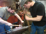
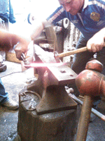
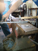
December 2010 saw Nigel's last clinic of the year, the most recent of many he has hosted at his forge for farriers who are interested in improving their skills in different areas of shoemaking. Chris Pell and Jason Evens attended this clinic, having attended others previously.
Tool and fullering was the day's forging venture. Nigel began by first checking the tooling blocks that Chris and Jason had; he made some adjustments to them so they worked more effectively by heating the entire blocks and then sinking in his own section to freshen them up before case hardening them. Once this was accomplished, Nigel moved on to demonstrating front tool and fullered hunter shoe and breaking down the stages of the importance of position and tool placement during the tooling procedure, and splitting the fullering phases. Nigel also pointed out how tool and fullering really is one of those arts in forging where through practise you develop a feel for it. After demonstrating the front and hind, both Chris and Jason had a go themselves with Nigel looking on and guiding them as necessary. Australian visiting farrier Michael Saunders – who is currently working alongside Nigel - acting as striker for them both! At lunchtime, Nigel's wife delivered the famous 'fish and chip' lunch, which always go down well at every clinic.
For the second part of the day, Nigel moved onto forging the tool and fullered hunter hind, breaking down the caulk and wedge forging moves and formation techniques. After this was done Chris and Jason embarked on the same with, once again, Nigel guiding and Michael Striking, burning even more calories and working off their lunch! With a little time spare at the end of the day, Jason asked Nigel to demonstrate a quick hind tool and fullered bar shoe off the hammer, specifically to show the formation of the scarfs and welding.
Both Jason and Chris did very well during the day with the shoes they produced from their new freshened blocks, they now certainly have strong basics of this art under their belts to practice and refine. Nigel wishes to thank Chris and Jason for attending and for the pictures they took of the day; Michael for being 'Conan' for the day, and to all the farriers that have attended any of Nigel's clinics over the past two years. With special thanks to Handmade Shoes UK for sponsoring some of these clinics, and his wife Teresa for providing catering for all the events.
New clinics planned for 2011 can be viewed at www.nigelfennell.co.uk; they are aimed at farriers wishing to prepare for higher examinations, assessments, embarking on competitions or wanting to refine and advance their personal forging skills. All the clinics are CPD recognised and participants gain 4 CPD points.
moreNEWS
morespecial offers
CPD Points Don't forget you can get 1 CPD point for writing a Farriery Related Article for Forge Magazine (max 4 points per year)
Attend a one day NAFBAE event for 4 CPD points, a half day is 3 CPD points and an evening event is 2 CPD points.
These include local meetings etc but exclude purely social events.
For further information contact the CPD office on 01773 341393.